
Vision Problems from Stroke and Head Injury
Our doctors have specialized in helping patients who have vision issues from stroke and brain injury for over 30 years.
According to the National Center for Health Statistics, there are over 8 million head injuries each year with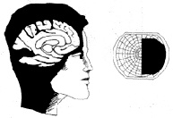 over 1.5 million of them classified as major injuries. The vast majority of people are saved by advances in modern medicine while only about 100,000 suffer fatal injuries. Another 500,000 people suffer from strokes each year, and it is estimated that there are over 3 million stroke survivors in the United Stated alone.
Millions of stroke and traumatic brain injury survivors suffer from visual problems. These visual problems range from dry eyes to visual field loss to double vision. Each case is different and the difficulty each patient has depends on the severity and location of the injury. Unfortunately, many patients with visual problems after a stroke or head injury fail to receive adequate vision rehabilitation.
Brain injury may affect vision in many ways. Below are some of the most common visual problems associated with stroke and traumatic brain injury.
Loss of Visual Field
A common visual effect of brain injury is the loss of one’s visual field or our ability to see to the side. There are many types of visual field losses that can occur, but the most common form is a homonymous hemianopsia or loss of half of the field of vision in each eye. If the posterior portion of the brain is damaged on one side of the brain, a loss of visual field occurs to the opposite side in both eyes. Patients often mistakenly believe the loss is just in one eye.
 Patients frequently bump into objects, and easily trip or fall over objects in their field loss. Going into crowded stores may become quite difficult, because people and objects suddenly appear in front of them from the blind side. Patients become afraid of leaving their homes and may even experience panic attacks. Additionally, the loss of visual field may also cause patients to miss words and have difficulty reading.
Patients with hemianoptic field loss may benefit from the Visual Field Awareness System developed by Dr. Daniel Gottlieb or the Peli Lens developed by Dr. Eli Peli, senior researcher at Harvard. Both of these systems increase the patient’s ability to detect objects on the side of their vision loss. Training the patient in scanning techniques is also an important part of the treatment.
Visual Spatial Disorders and Visual Neglect
Patient may experience a variety of visual spatial disorders. When certain portions of the brain are damaged, the patient may fail to appreciate space to one side, which is usually to the left. Unlike visual field loss, this problem is not a physical loss of sensation, but rather a loss of attention to the area. A man with neglect may no longer shave one side of his face. Patients with visual neglect have more difficulties than those with only visual field loss. Unfortunately, both neglect and field loss may occur together. Other visual-spatial disorders may occur as well. Patients may experience difficulty navigating themselves even in familiar areas. Patients also misjudge the straight-ahead position and can confuse right versus left.
Vertigo, Dizziness and Impaired Eye Movements
Smooth and accurate eye movements are essential in reading, tracking objects and compensating for body movements. Following injury to the brain, movements may become more jerky in nature. As we move or tilt our head, our inner ears sense the angle at which we are tilting and causes compensatory eye movements. After head injury, these compensatory movements may become impaired. Nystagmus, a jerky motion of the eyes, may also occur. When acquired later in life, nystagmus results in a vertigo-like sensation or a feeling that the world is moving. Damage to the brain stem often results in dizziness.
Double Vision
Our eyes must point precisely at the same point in space to prevent diplopia or double vision. Each eye has six external muscles that move the eyes together as a team. If control is impaired to one or more muscles, the eyes cannot maintain alignment in all positions of gaze. This may occur due to damage to the control centers for the III, IV and VI cranial nerves. Double vision may be constant or intermittent. The patient may experience normal single vision in the straight ahead position, but suddenly have double vision on looking to one side.
Eyestrain and Difficulty in Reading
As we bring reading material close to our eyes, both eyes must turn in together as a team. It is called ocular convergence. This ability is frequently impaired in brain injury resulting in fatigue or discomfort while reading. Orthoptic therapy and prism lenses may aid this problem.
After injury, younger patients may experience more problems focusing at near. This may present simply as difficulty in reading. This is usually due to damage to the oculomotor nerve (CN III). This nerve is responsible for controlling the eye’s ability to focus by changing the shape of the crystalline lens. Patients benefit from the use of bifocals to help compensate.
Eye movements called saccades are used to jump from word to word as we read. Impairments in saccades may result in difficulty reading smoothly along a line of print. Visual field loss may also impair reading. When the loss of field borders on the central area of the retina used in reading, patients may lose their place or have difficulty in reading long words. Impairments in cognitive skills and memory may also limit reading. Some patients may acquire an alexia, a loss of reading ability. Therapists may be able to rebuild reading skills in some patients. For those still unable to read, electronic scanner/reading machines like the Kurweil Omni system can read to the patient.
Light Sensitivity
Light sensitivity is quite variable. Some patients experience no problems while others have severe light sensitivity. Much like the volume control on a radio being broken, patients seems to have difficulty adjusting to the various lighting levels. Tinted eyewear, especially amber filters, may aid the patient and light sensitivity may improve with treatment of other vision problems.
Dry Eyes
Dry, burning or gritty eyes may occur after brain injury. It may result from a decrease in the blink rate, or poor closure of the lids. Artificial tears or tear duct plugs will usually control the problem
Visual Hallucinations
Visual hallucinations may be formed objects such as a person or figure or may be unformed such as flashes of lights, stars or flickering distortions.
Impaired Visual Memory
Memory is often impaired after stroke or head injury. In rare cases very specific types of memory processing is impaired. A patient may no longer be able to recognize faces, objects or letters.
Low Vision Care
The visual problems of acquired brain injury may affect nearly all aspects of vision and can hinder normal recovery. Early vision evaluation is crucial. A clinician skilled in both low vision and brain injury is often needed to understand the interaction of all of these visual problems in order to make the appropriate low vision rehabilitation plan for each patient with acquired brain injury. The long road back from brain injury requires the teamwork of many doctors and therapists and most of all time and patience throughout the rehabilitative process.
Hemianopsia.Net www.hemianopsia.net
Our doctors have worked with stroke and traumatic brain injury patients for over 25 years. This website built by our doctors discuss all the aspects of hemianopsia and treatment options available. Our doctors use special prismatic eyewear to shift the image into the patient's remaining visual field to help the patient get a better understanding of their position and whereabouts.